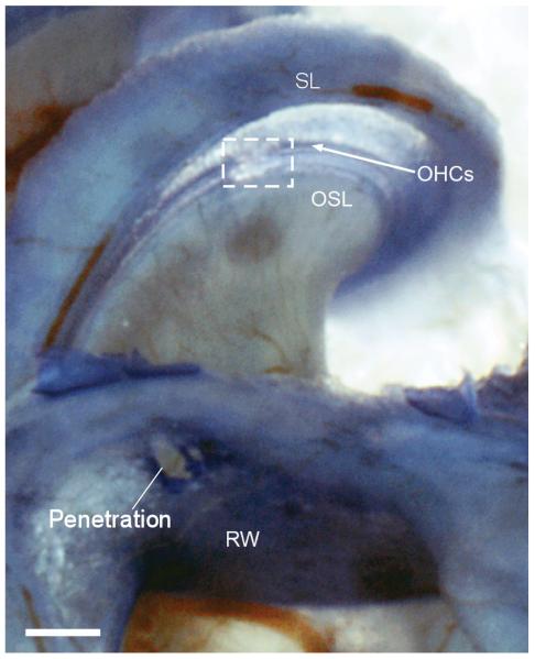
Figure 1.
Example of damage to basilar membrane caused by the electrode. The preparation shown is a decalcified toludine blue stained whole mount. Damage is outlined by the box. The physiology data in Figures 2-4 is from this case. SL: spiral ligament; OHCs: Outer hair cells; OSL: osseous spiral lamina; RW: Round window.

