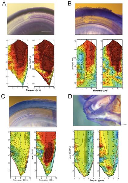
Figure 6.
A: Extensive damage on the basilar membrane; the loss of electrophysiologic activity was extensive and irreversible. The damage consisted of a clear breach of the basilar membrane, loss of hair cells, and damage to supporting cells lateral to the OHCs. B: Trauma similar to that previously seen in the case shown in Figures 1-4. The damage consisted of a clear breach of the basilar membrane, loss of hair cells and some supporting cells. The loss of response in the CAP was extensive but that to the CM was moderate. C: Damage on the basilar membrane restricted to a small site underneath the hair cells and tunnel of Corti. On close inspection there appeared to still be a thin layer of membrane separating the scala tympani and scala media. Although there was loss of hair cells, it is possible that the reticular lamina was either not breached such that mixing of endolymph and perilymph either did not occur or was minimal. The magnitude of response loss was moderate for the CAP, and minimal for the CM. D: No damage to the basilar membrane. Instead, there was a region of damage on the OSL, which appeared to be a flake of bone that was lifted up allowing stain to be trapped. In this case, the damage to both the CM and CAP was minimal, and reversed to a substantial degree after withdrawal of the electrode.

