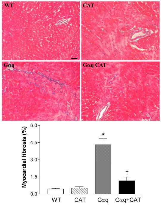
Figure 5.
Representative photomicrographs of Masson Trichrome staining for fibrosis in left ventricular myocardium of wild-type (WT), catalase (CAT), Gαq and Gαq/CAT mice at 20 weeks lf age. Increased myocardial fibrosis was present in Gαq mice, and was inhibited in Gαq/CAT mice. Myocardium stains red, and collagen stains blue. The bar in the left upper panel indicates 50 μm.

