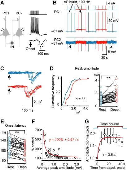
Figure 1. Modulation of slow (late-onset) disynaptic IPSP by presynaptic somatic V m.
(A) Left, schematic diagram of the PC-PC paired recording (IN indicates those unidentified inhibitory interneurons that mediate the disynaptic IPSPs). Right, an AP burst (15 APs at 100 Hz) evoked by a train of current injection in PC1 induced a disynaptic response in PC2 with a long latency from the onset of the AP burst. * indicates individual IPSPs. (B) Example recording showing that presynaptic depolarization increased the amplitude of AP burst-induced disynaptic IPSPs. (C) Overlay of the IPSPs evoked at resting (blue) and depolarized (red) V m in the presynaptic PC. Arrows indicate the onset of the AP train. Notice that presynaptic depolarization caused a reduction in failure, increased the amplitude, and shortened the latency of the disynaptic IPSPs. (D) Left, cumulative frequency distribution of the tested connections (n = 38 PC-PC pairs) by the average amplitude of disynaptic IPSP at resting (blue) and depolarized V m (red); right, pooled results showing changes of the average amplitude at the two V m levels in individual PC-PC pairs. (E) Pooled results (n = 38 pairs) showing that the onset latency of IPSPs was shortened by presynaptic depolarization. (F) The percentage increase was dependent on the average amplitude of disynaptic IPSPs (n = 38 pairs). Red line, hyperbolic fit. (G) Average time course of the facilitation in PC-PC pairs that showed significant increase in IPSP amplitude (n = 12 pairs tested). Error bars represent s.e.m. ** p<0.01.

