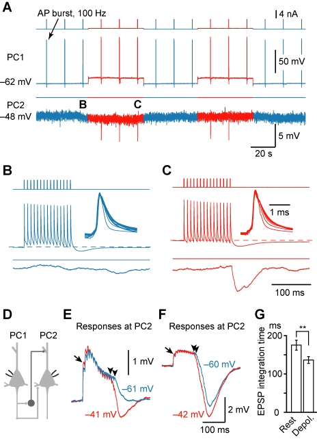
Figure 2. Presynaptic depolarization turns on recurrent inhibition and shortens the integration time of EPSPs.
(A) Example PC-PC paired recording showing that the disynaptic IPSP only occurred at a depolarized V m. (B and C) Parts in (A) were expanded for clarity. Insets, overlay of the somatic APs during the train indicating that no AP failure occurred. (D) Schematic drawing of the recordings (for E–G) from PC-PC pair that had both the monosynaptic excitatory connections and the disynaptic inhibitory connections. (E) An example showing that disynaptic IPSP (average of 33 trials), which occurred only when the presynaptic V m was depolarized, shortened the EPSP summation time (arrowheads). The arrow indicates a facilitated EPSP. (F) Similar example (average of 15 trials) as shown in (E) except that disynaptic IPSP occurred at both depolarized and resting presynaptic V m. Note the difference in the time window of EPSP summation (arrowheads). (G) Group data (n = 9 PC-PC pairs) indicated that presynaptic depolarization shortened the time window for EPSP integration. ** p<0.01.

