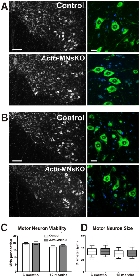
Figure 2. ß-actin is not required for motor neuron viability in vivo.
(A) Representative images of Nissl stained spinal cord sections from the lumber enlargement of 6 and (B) 12 month old mice. Magnified images at right show no indication of chromatolysis in motor neurons. DAPI staining is in blue. Scale bar 100 µm left, 30 µm right. (C) Quantification of motor neurons per section at 6 and 12 months of age (n = 3 for each genotype per time point). No statistically significant differences were observed between control and Actb-MNsKOs at either time point. (D) Quantification of motor neuron size at 6 and 12 months of age. Data plotted as mean ± standard error of the mean.

