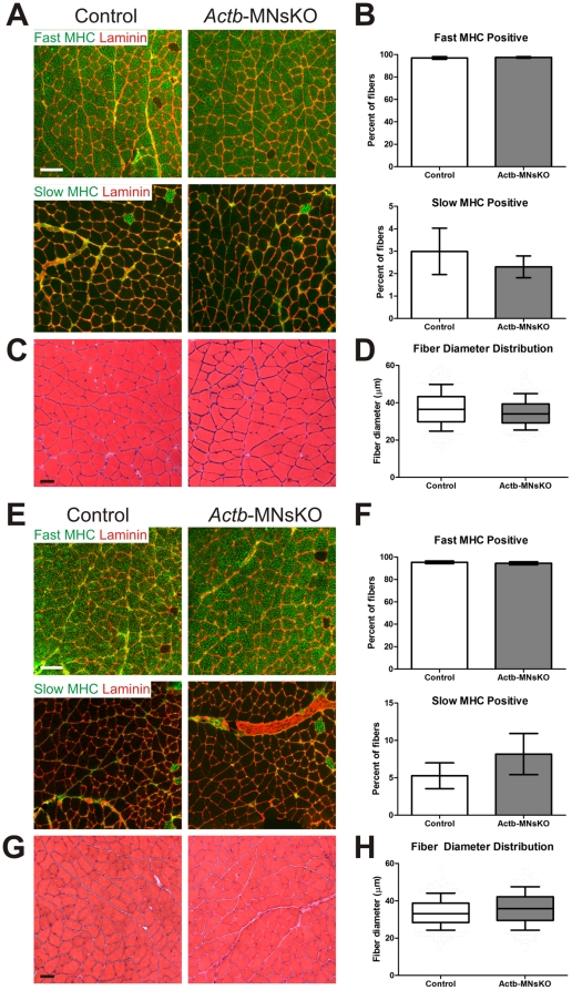
Figure 5. Histological analysis of skeletal muscle in Actb-MNsKO mice.
Fiber type composition and distribution was assessed in the gastrocnemius muscle of 6 and (A–B) and 12 month old (E–F) control and Actb-MNsKO mice. Antibodies to fast and slow myosin heavy chain were used to identify fast and slow twitch fibers respectively. Anti-laminin staining was used to delineate the borders of muscle fibers. Scale bar 100 µm. Fiber diameter was analyzed from hematoxylin and eosin stained sections of the gastrocnemius muscle at 6 (C) and 12 months (G) of age. Scale bar 50 µm. Box and whisker plots of fiber diameter distribution at 6 (D) and 12 (H) months of age.

