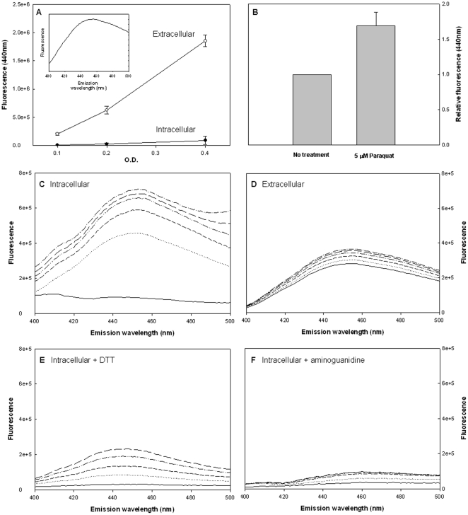
Figure 1. Kinetic studies of AGEs formation and secretion.
Bacterial growth and sample collection were described in materials and methods. AGEs-specific fluorescence (Ex. 370, Em. 440) was determined and normalized to cells density. A) AGEs-specific fluorescence during growth in the intracellular and extracellular fractions. The insert shows the distribution of AGEs with excitation at 370 nm. B) Effect of 5 µM paraquat on Extracellular accumulation of AGEs. C–D) Kinetics of AGEs formation in vitro. Bacteria were separated from extracellular fractions and sonicated (intracellular fraction). C) The intracellular and D) extracellular fractions were incubated at 37°C and AGEs-specific fluorescence was determined every 30 minutes (Ex. 370, Em. 440–500). Effect of E) 2 mM DTT or F) 50 mM aminoguanidine on AGES in the intracellular fractions. All data represent three independent experiments, in Figures C–F a representative result is shown.

