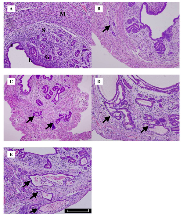
Figure 1.
HE staining of paraffin-embedded sections. A: Uterus of 140 ± 5 d ICR mice as control. B:Uterus of 90 ± 5 d ICR mice with biopsy confirmed adenomyosis; C: Uterus of 140 ± 5 d ICR mice with biopsy confirmed adenomyosis; D: Uterus of 190 ± 5 d ICR mice with biopsy confirmed adenomyosis; E: Uterus of 240 ± 5 d ICR mice with biopsy confirmed adenomyosis; (L, luminal epithelium; G, glandular epithelium; S, stromal cell; M, myometrium cell; arrows, ectopic endometrium; Bar = 50μm).

