
Figure 1.
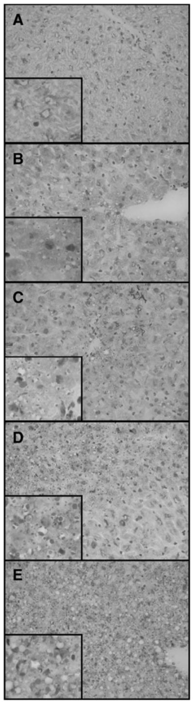
Liver microvesicular scoring of Oil-Red-O staining. Liver sections showing the degrees of microvesicular steatosis as demonstrated by the presence of positive red staining fat droplets within the hepatocyte cytoplasm. No microvesicular steatosis identified (A). Minimal microvesicular steatosis with rare small focal areas involving periportal areas (B). Mild microvesicular steatosis with multifocal areas involving periportal areas (C). Moderate microvesicular steatosis involving zones periportal or mid-zonal regions and/or with areas of portal to portal bridging microvesicular steatosis (D). Severe microvesicular steatosis with diffuse steatosis involving all zones of the hepatic parenchyma (E). Images are at ×400 and insets are at ×1000.