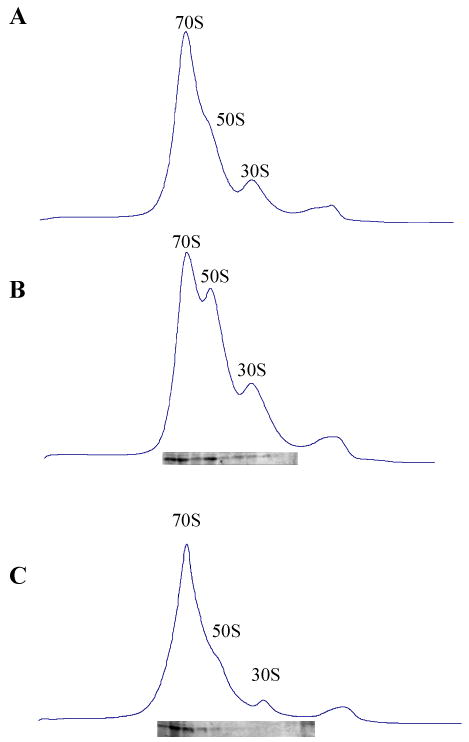
Fig. 7. Effect of Paromomycin on RatA activity in vitro.

70 pmol of 70S ribosomes were incubated in the buffer S at 30°C for 10 min, then 0.4 μg of RatA or 400 μg/ml of paromomycin was added, the mixture was incubated for another 10 min, then the concentration of Mg2+ was increased to 6 mM and the mixture were further incubated for 10 min before loading on to a 5%–40% sucrose density gradient in buffer T following ultracentrifugation and the sedimentation behavior was monitored using spectrophotometer. (A) Ribosome profile in the presence of 6 mM Mg2+. (B) Ribosome profile in the presence of 6 mM Mg2+ and 0.4 μg of RatA. (C) Ribosome profile in the presence of 6 mM Mg2+, 0.4 μg of RatA and 400 μg/ml of paromomycin. In B and C, Western blot analysis was carried out to detect RatA in each gradient fraction using anti-RatA antibody.
