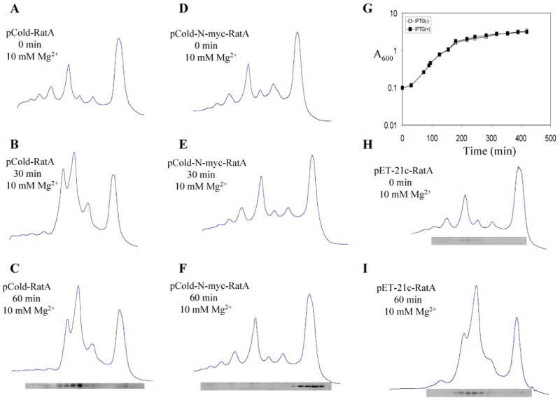
Fig. 8. Addition of N-terminal myc-tag to RatA eliminated its toxicity.

Ribosome profiles were analyzed in 10 mM Mg2+ by sucrose density gradient centrifugation as described in the Experimental Procedures. (A) Polysome profiles of E. coli BL21(DE3) containing pCold-RatA without RatA induction, (B) at 30 min after RatA induction, (C) at 60 min after RatA induction. (D) Polysome profiles of E. coli BL21(DE3) containing pCold–N-myc-RatA without RatA induction, (E) at 30 min after RatA induction, (F) at 60 min after RatA induction. (G) Growth curve of E. coli BL21(DE3) containing pCold–N-myc-RatA. (H) Polysome profiles of E. coli BL21(DE3) containing pET-21c-RatA without RatA induction and (I) at 60 min after RatA induction. In C, F, H and I, Western blot analysis was carried out to detect the RatA protein in each gradient fraction using monoclonal anti-poly-histidine antibodies.
