
Fig. 3.
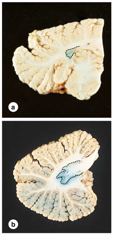
Gross appearance of the DN in FRDA. (a) FRDA; (b) normal control. The interrupted lines indicate the approximate location of the DN gray matter. The small size of the nucleus in FRDA is particularly apparent on a macrostain for iron (a). The normal DN shows the typical meandering gray matter ribbon (b). Iron reaction product does not co-localize with the gray matter but extends into the white matter of hilus and fleece.