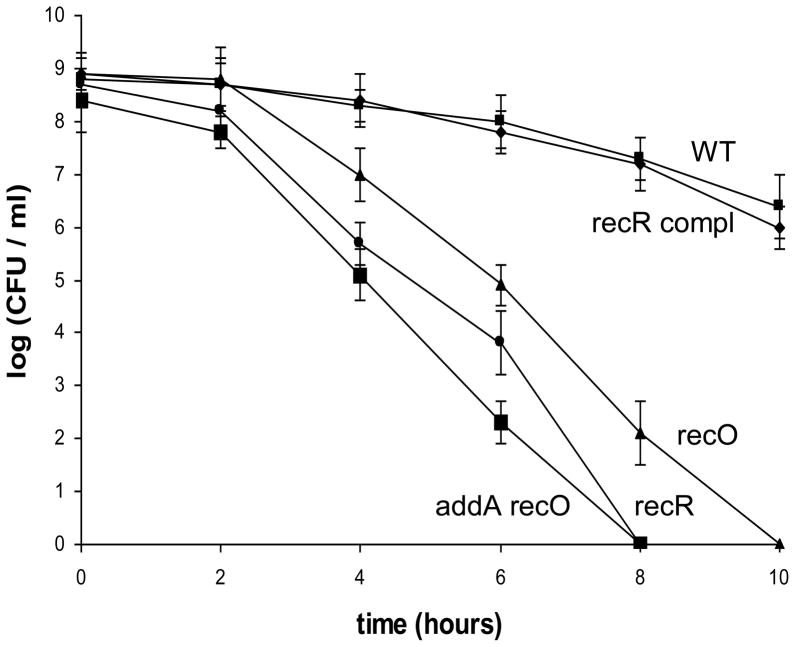
Fig. 1. Survival of H. pylori cells upon exposure to air.
H. pylori cell suspensions in PBS were incubated at 37°C under normal atmospheric conditions (21% partial pressure O2, and no alteration of CO2 partial pressure). Samples were removed at the times indicated in the x axis and were used for plate count determinations in a 5% oxygen environment. The data are the means of three experiments with standard deviation as indicated. Symbols: square, wild type; diamond, recR complementation strain; triangle, recO::aphA; circle, recR::cat; large square, addA::cat recO::aphA. Based on statistical analysis (Student t-test), the cell survival differences between the WT and the mutant strains are significant (P<0.01) for all the data points except for the 2 h time point.

