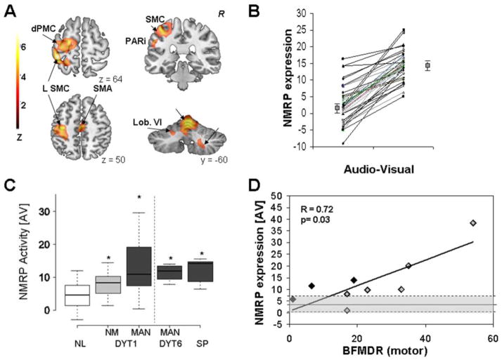
Figure 3. Normal motor-related activation pattern (NMRP).
A. NMRP as identified by ordinal trends canonical variates analysis (OrT/CVA) of 78 H215O PET scans from 18 healthy volunteers (Carbon et al., 2010). [Color scale: Positive voxel weights thresholded at Z=3.09, corresponding to regions that contributed significantly (p<0.001) to network activity. The displayed voxel weights were found to be reliable (p<0.0001) on bootstrap estimation]. B. NMRP expression in the subjects comprising the original derivation sample. For all subjects and runs, pattern expression increased during the performance of the motor task (p<0.0001, (Carbon et al., 2010)). C. NMRP scores for manifesting (MAN) and non-manifesting (NM) DYT1 carriers and controls (left panel), and for MAN DYT6 carriers and subjects with sporadic (SP) primary dystonia (right panel). For all subjects, the network values were measured in H215O PET scans acquired in the audio-visual (AV) condition (Carbon et al., 2010). In this non-motor (“sensory”) condition pattern expression was elevated in both groups of mutation carriers and in the SP subjects. [Significant post-hoc comparisons with controls (p<0.05) are denoted by asterisks]. D. NMRP expression in MAN DYT1 carriers measured in the audio-visual (AV) condition correlated with clinical severity ratings according to the Burke-Fahn-Marsden Dystonia Rating Scale (BFMDRS). [Black diamonds: subjects without contractions at rest; gray diamonds: subjects with constant or with occasional contractions at rest. The normal mean and range are indicated by shading].
[A-D: Adapted from Brain, Increased sensorimotor network activity in DYT1 dystonia: a functional imaging study, 690-700, Copyright 2010, with permission from Oxford University Press]

