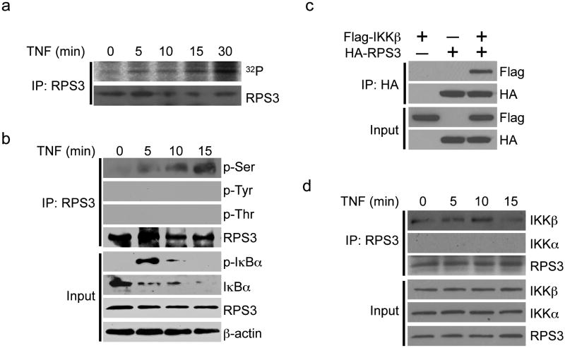
Figure 1. RPS3 is phosphorylated and associates with IKKβ in response to NF-κB activation.
(a) 32P-labeling assays were performed with HEK 293T cells stimulated with TNF (20 ng/ml) for the indicated times. Whole-cell lysates were subjected to immunoprecipitation (IP) with RPS3 antibody, followed by autoradiography or immunoblotting with RPS3 antibody. (b) Whole-cell lysates (Input) from Jurkat cells stimulated as indicated were directly immunoblotted for indicated proteins, or serine-phosphorylated (p-Ser), tyrosine-phosphorylated (p-Tyr), or threonine-phosphorylated (p-Thr) proteins, or RPS3 after immunoprecipitation with RPS3 antibody (IP: RPS3). (c) Interaction between HA-RPS3 and Flag-IKKβ in HEK 293T cells assessed by immunoprecipitation and immunoblot (IP: HA). (d) Whole-cell lysates (Input) from Jurkat cells stimulated as indicated were directly immunoblotted, or after immunoprecipitation (IP) with RPS3 antibody, for IKKα, IKKβ, or RPS3. Data are representative of at least two independent experiments.

