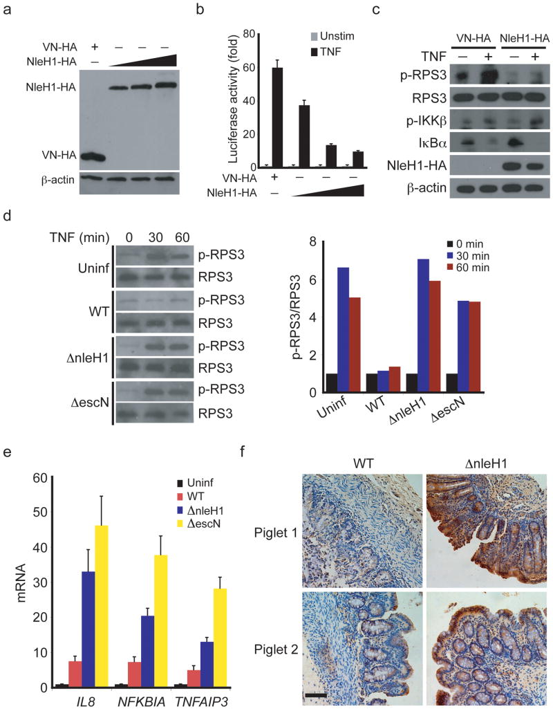
Figure 6. The bacterial effector protein NleH1 blocks RPS3 S209 phosphorylation.
(a) 293T cells were transfected with control VN-HA or NleH1-HA plasmids and whole cell lysates were derived and immunoblotted for HA and β-actin as a loading control. (b) NF-κB luciferase assay (mean and s.d., n = 3) using 293T cells transfected with control VN-HA or NleH1-HA plasmids together with a 5 × Ig κB site-driven luciferase reporter gene. (c) 293T cells overexpressing either vehicle protein (HA) or NleH1-HA were stimulated with (+) or without (-) 50 ng/ml of TNF for 15 min. The derived whole cell lysates were immunoblotted for S209 phosphorylated RPS3 (p-RPS3) and indicated proteins. β-actin served as a loading control. (d) HeLa cells were left uninfected (Uninf) or infected for 3 h with wild-type (WT) E. coli O157:H7 or strains with isogenic deletions in the escN (ΔescN) or nleH1 (ΔnleH1) genes, followed by TNF treatment for the indicated periods. Whole cell lysates were extracted and immunoblotted with antibodies specific for normal RPS3 or S209 phosphorylated RPS3 (left). Densitometry of all bands was performed, and the intensity of each p-RPS3 band was normalized to corresponding RPS3 band. The fold change of p-RPS3/RPS3 was further normalized to the 0-min samples (set as 1.0) in cells infected with the indicated E. coli O157:H7 strains (right). (e) Transcript abundance relative to uninfected cells assessed by RT-PCR analysis of HeLa cells infected for 3 h with E. coli O157:H7 strains as in (d). The relative mRNA abundance of IL8, TNFAIP3, and NFKBIA were normalized to GAPDH expression (mean and s.d., n = 3). (f) Immunohistochemistry for S209 phosphorylated RPS3 in paraffin-embedded piglet colons derived from gnotobiotic piglets infected with E. coli O157:H7 EDL933 strains possessing (WT) or lacking NleH1 (ΔnleH1), using phospho-RPS3 antibody and 3,3′-diaminobenzidine as a substrate (brown). Nuclei were counterstained with hematoxylin (blue). Size bar represents 25 μm. Representative images from two piglets are shown. Data are representative of two (a, c), four (b), three (d, e), and six (f) independent experiments, respectively.

