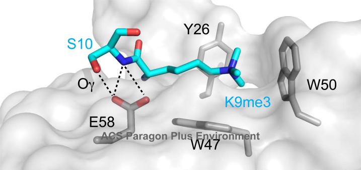
Figure 4.
Depiction of H3K9me3 and S10 side chains in the Pc-H3K27me3 structure, with Pc depicted in surface representation. Polar contacts made between the side chain of Pc E58 and H3S10 (backbone nitrogen in blue, side chain oxygen in red and denoted Oγ) illustrate how phosphorylation at S10 could disrupt the interaction. Aromatic cage residue side chains are also shown in stick representation.

