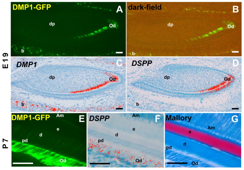
Figure 3. Expression of DMP1-GFP transgene in developing mandibular incisors.
Epifluorescence (A, E), dark-field (B) and pseudo-colored bright-field (C, D, F) images of sections through developing incisor teeth at E19 (A–D) and P7 (E–G). In each stage of development, adjacent sections were processed for different analyses. (A–D) At E19 DMP1-GFP is not expressed in apical regions containing pre-odontoblasts and is detected in the incisal region containing secretory/functional and fully differentiated odontoblasts expressing DSPP (C) and DMP1 (D). DMP1-GFP is expressed at higher intensity in fully differentiated as compared to secretory odontoblasts. DMP1-GFP is expressed in the osteoblasts and osteocytes of the developing alveolar bone (indicated as b) that were expressing DMP1, but not DSP.
(E–G) are images through incisors at P7. Intensity of DMP1-GFP expression increased with further odontoblast differentiation during postnatal life and extended into the odontoblast processes (E–F). G represents an image of a section stained with Mallory. Abbreviations: Am, ameloblast; B, bone; d, dentin; dp, dental pulp; E, enamel; Od, odontoblast; pd, predentin. Scale bar in all images=100mm.

