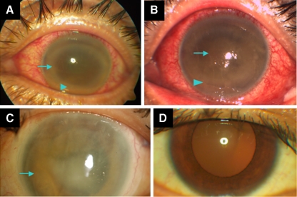
Fig. 1.
a–d External photographs. a Left eye, at presentation, dark corneal ring infiltrate (arrow), and brown hypopyon (arrowhead). b Four days later, progression of dark hypopyon (blue arrowhead) and development of a brown papillary membrane (arrow). c Left eye, postoperative corneal edema and significant iris heterochromia (arrow). d Right eye, showing the patient's original iris color, obtained at a similar time point as c

