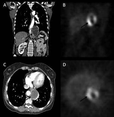
Figure 1.

Coronal (A) and axial (C) CT images demonstrating a 65×65×95 mm partly solid partly cystic mass situated in the posterior mediastinum. The mass is predominantly occupying the left retrocrural space extending along the vertebral bodies of T10–L1 abutting the oesophagus, aorta and diaphragm. 123I-MIBG-SPECT (ioflupane-metaiodobenzylguanidine-single photon emission CT) images at a position that correlates with the mass demonstrated on CT (B) and (D). The peripheral isotope uptake correlates to the solid elements of the tumour, whereas the central lack of uptake correlates to the cystic elements.
