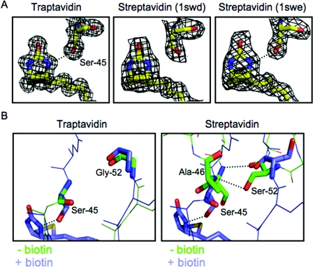
Figure 4. Hydrogen bonding by Ser45 in Tr.
(A) The electron density for biotin and Ser45 surrounds a stick model of the atoms involved, for Tr (left-hand panel) and two different streptavidin structures (1SWD in the middle panel and 1SWE in the right-hand panel). Hydrogen bonds are indicated by a broken line. (B) The rival hydrogen bonding by Ser45. An overlay of the L3/4 region, with key residues highlighted in stick format. For Tr (left-hand panel) with and without biotin L3/4 is closed, with no hydrogen bond between Ser45 and Gly52. For SA (right-hand panel) without biotin and with an undefined L3/4 (1SWC, chain B), Ser52 forms hydrogen bonds to Ser45 and Ala46 (indicated by broken lines). When biotin is bound and the loop is closed (1SWE, chain D), Ser45 now forms a hydrogen bond to biotin.

