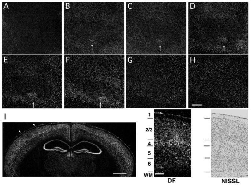
Figure 2.

Activity-dependent cpg15 expression is localized to layers II/III and IV of barrel cortex. Representative dark-field photomicrograph of in situ hybridization for cpg15 mRNA on serial tangential sections through flattened cortex of a 4-week-old mouse after 12 h single whisker experience. The sections progress from layer I (A) to layer VI (H). Arrows mark the cortical region representing the spared whisker in layers II through IV (B–F). Scale bar, 0.5 mm. (I) cpg15 mRNA localization is shown in a coronal section through the brain of a mouse treated as above. The contralateral hemisphere to the spared whisker shows cpg15 up-regulation in the region representing the D1 whisker, with a concomitant down-regulation in the surrounding barrel field. White arrowheads indicate the region of the barrel field shown at high magnification on the right. Scale bar, 1 mm. Dark-field view shown side by side with a bright-field view of an adjacent Nissl stained section delineating cortical layers. Scale bar, 0.1 mm.
