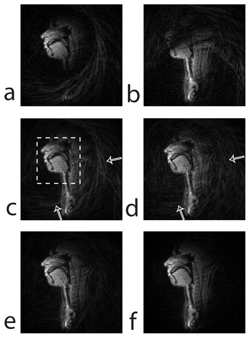
Figure 3.

Midsagittal images with a large reconstruction field-of-view (FOV) of 38 × 38 cm2 reconstructed from the data acquired in static posture. (a–c) Conventional bit-reversed 13-interleaf UDS. (a) image from coil 1, (b) image from coil 2, (c) root sum-of-squares (SOS) of the coil 1 and coil 2 images. The region within the dashed box in (c) is the vocal tract regions of interest (ROIs). For speed-up of the spiral acquisition, the FOV of the 13-interleaf UDS is typically chosen to be small such that aliasing artifacts are not observed in the vocal tract ROIs. (d–f) Root SOS of the coil 1 and coil 2 images reconstructed from data acquired using the spiral golden-ratio acquisition: reconstruction from (d) 13-TR, (e) 21-TR, and (f) 34-TR data. Spatial aliasing artifacts are completely removed in (f) because of larger FOV available from the golden-ratio method. The image SNR in (f) is higher than that in (d) and (e).
