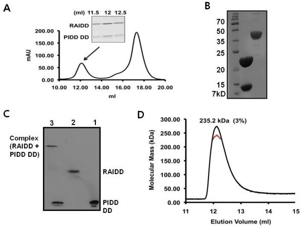
Figure 5. Complex formation of full-length RAIDD and PIDD DD, binary PIDDosome complex, at low salt.

A. Separately purified both full-length RAIDD and PIDD DD are mixed together in the buffer containing 20 mM Tris-HCl pH 8.0, 50 mM NaCl, and 1 mM DTT and incubated R.T. for 1 hour. Gel-filtration profile showing formation of the complex between full-length RAIDD and PIDD DD. SDS-PAGE of gel filtration fractions showing formation of the complex. B. The final concentrated complex peak containing both RAIDD and PIDD showing on SDS-PAGE. The complex eluted at around 12 ml was collected and concentrated to 6 mg/ml: lane 1, final complex sample. Lane 2, Apo AI protein for comparison of concentration. C. Native-PAGE analysis of the interaction between full-length RAIDD and PIDD DD: lane1, PIDD DD only; lane 2, full-length RAIDD only; lane3, full-length RAIDD mixed with PIDD DD. D. Multi-angle light scattering (MALS) measurement of the RAIDD: PIDD DD complex peak, showing the molecular mass of the complex.
