
Figure 1.
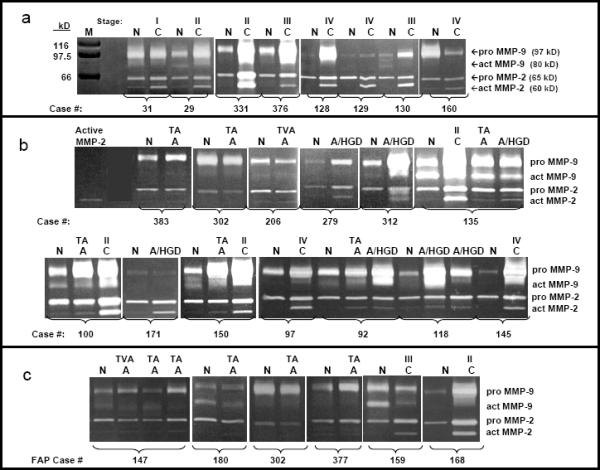
1a. MMP-2 and MMP-9 activities in 8 matched pairs of normal and cancer tissues. Gelatin zymograms for eight pairs of patient-matched normal mucosa (N) and colorectal cancer (C) extracts with Coomassie blue stained protein markers (M) of defined molecular mass to the left. Active MMP-2 was detected as a 60 kD band showing stronger activity in all cancer samples compared to matched normal mucosa. Pro-MMP-2, detected as a 65 kD band, was present in all samples, with higher activity in 6/8 cancers compared to matched normal mucosa. Active MMP-9 activity was detected as a weak 80 kD band in two normal mucosa samples (N29 and N130). Pro-MMP-9 activity was detected as a 97 kD band demonstrating a wide range of variation in activity levels from almost non-detectable (N129) to very high levels (C331) and increased activity in 5/8 cancer samples compared to normal mucosa. Cancer stage is designated above each cancer tissue (Stage I, S I; Stage II, S II; Stage III, S III; Stage IV, S IV).
1b. MMP-2 and MMP-9 activities in 13 matched sets of normal, adenoma without and with high grade dysplasia and cancer tissues. Gelatin zymograms for sets of normal mucosa tissues (N) with patient-matched adenomas without HGD (A) and/or adenomas with high grade dysplasia (A/HGD) and/or colorectal cancer (C) with purified active MMP-2 standard in the far left lane. All samples with the same case number were removed simultaneously from a given individual. Adenomas without HGD (A) are also designated as TA (tubular adenoma), TVA (tubulovillous adenoma) or VA (villous adenoma). Cancer samples (C) are also designated by stage, as described for Fig 1a. The 60 kD active MMP-2 activity was very low or not detectable in a majority of normal mucosa extracts, despite clear detection of pro-MMP-2 and pro-MMP-9. Higher levels of active MMP-2 activity were detected in many colorectal adenomas without HGD, adenomas with HGD, and cancer compared to normal. Changes in active MMP-2 levels in different tissues removed at surgery from a single subject (Cases 92, 100, 135 and 150) illustrate a step-wise increase in active MMP-2 activity with progression from normal colorectal mucosa to adenoma without HGD to adenoma with HGD to carcinoma. Levels of 65 kD pro-MMP-2 activity, while present in normal mucosa at much higher levels than active MMP-2, did not change much in colorectal adenomas, with or without high grade dysplasia, but did increase in most carcinomas. Pro-MMP-9 activity levels varied dramatically from case to case, being higher in some adenomas and cancers than matched normal (Cases 92, 100, 135, 145, 150, 279 and 312), showing minimal changes by tissue type in other cases (Case 171) and were sometimes lower in adenoma or cancer compared to normal (Cases 206 and 302).
1c. MMP-2 and MMP-9 activities in normal and adenoma tissues from 4 cases of FAP and in normal and cancer tissues from 2 cases of attenuated FAP. Gelatin zymograms for sets of normal mucosa tissues (N) with patient-matched adenomas without HGD (A) and/or colorectal cancer (C). Adenomas without HGD (A) are also designated as TA (tubular adenoma), TVA (tubulovillous adenoma) or VA (villous adenoma). Cancer samples (C) are also designated by stage, as described for Fig 1a. The 60 kD active MMP-2 activity was very low or not detectable (Cases 180, 302, 377 and 2 of 3 adenomas from Case 147) or very low (1 adenoma from Case 147) in all adenomas. Higher levels of active MMP-2 activity were detected in the 2 cancers from cases 159 and 168 compared to normal. Levels of 65 kD pro-MMP-2 activity also did not change much in colorectal adenomas (Cases 180, 302 and 2 of 3 adenomas from Case 147) but did increase in both carcinomas compared to normal. Pro-MMP-9 activity levels again varied being higher in some adenomas and cancers than matched normal (Cases 147, 377 and 168) with minimal changes by tissue type in other cases (Case 180) and were sometimes lower in adenoma or cancer compared to normal (Cases 302 and 159).