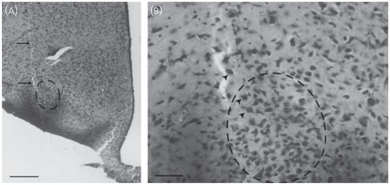
Fig. 2.

(A) Photomicrograph of a Nissl-stained section depicting the infusion tract (arrows) from a female infused with 60 ng of dopamine-β-hydroxylase-saporin into the ventrolateral division of the ventromedial hypothalamus. Scale bar = 500 μm. (B) Infusions caused minimal damage, and neurones at the infusion site (arrowheads) exhibited a normal distribution and morphology. Scale bar = 100 μm.
