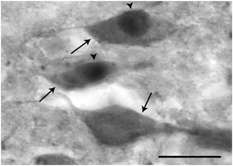
Fig. 3.

Photomicrograph of dopamine-β-hydroxylase-immunoreactive (DBH-IR) and DBH/Fos-IR cells in the rostral A2 nucleus. DBH-IR cells (arrows) were identified by their brown cytoplasmic staining. Dark purple-black nuclear label was observed in Fos-IR cells (arrowheads). Single- and double-labelled cells were readily identifiable using a 20× objective. Scale bar = 20 μm.
