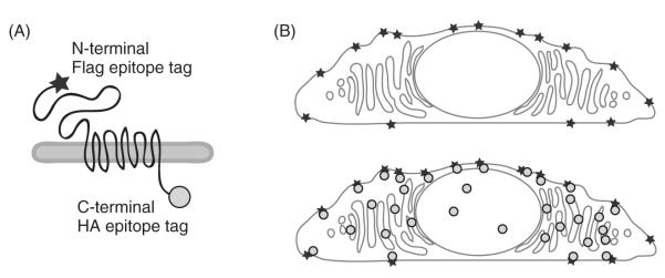
Fig. 1.
Surface immunofluorescence detects the pool of polycystin-1 (PC1) at the plasma membrane. The N-terminal Flag epitope tag on PC1 is marked with one antibody (represented by a star), while the intracellular C-terminal HA epitope tag is detected with a second antibody (represented by a circle) (A). The protocol for surface immunofluorescence is optimized to mark only the PC1 that has reached the plasma membrane (B, upper panel), while adding the HA antibody after permeabilizing the cells allows a visualization of the total amount of PC1 expressed in the same cell (B, lower panel).

