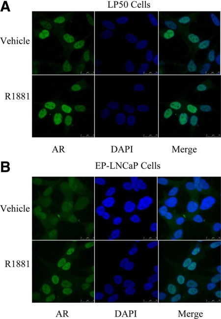
Fig. 2.
Subcellular localization of AR in LP50 cells and in EP-LNCaP cells. LP50 cells (A) or EP-LNCaP cells (B) grown in chamber slides were depleted of hormones and then treated with either vehicle or R1881 for 12 h. Immunofluorescence staining for AR was performed using a primary rabbit antibody to AR and a bovine antirabbit IgG-FITC as the secondary antibody. The nuclei were stained with 4′,6-diamidino-2-phenylindole (DAPI), and fluorescence images were captured by confocal microscopy. A, Representative images show predominant nuclear localization of AR (green fluorescence) in LP50 cells both with and without R1881 treatment. B, Representative images show a mixed distribution of AR (green fluorescence) between nuclear and cytosolic compartments in the absence of hormone but a predominantly nuclear distribution after R1881 treatment. Note that the brighter fluorescence staining of AR in both A and B upon R1881 treatment is consistent with the expected stabilization of AR by R1881.

