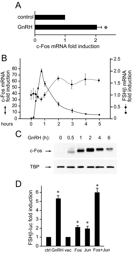
Fig. 1.
c-Fos induction by GnRH precedes induction of FSHβ. A, Mouse primary pituitary cells were dispersed in culture and treated with a 5-min pulse of vehicle of 10 nm GnRH. mRNA was extracted 1 h later and subjected to quantitative RT-PCR. c-Fos mRNA concentration was normalized to GAPDH mRNA in the same sample and then to the ratio in vehicle-treated cells. B, LβT2 cells were treated with GnRH for different lengths of time, after which the mRNA was extracted and the amount of c-Fos and FSHβ mRNA was analyzed by real-time quantitative RT-PCR. C, Whole-cell extracts from LβT2 cells treated with GnRH, time indicated above each lane, were subjected to Western blots for c-Fos and TATA-binding protein (TBP) as loading control. D, LβT2 cells transiently transfected with a 1-kb mouse FSHβ-luciferase reporter plasmid were treated for 5 h with 10 nm GnRH or cotransfected with expression vectors for c-Fos and c-Jun (Fos, Jun) or empty vector control (vec), as indicated. The luciferase/β-gal normalized values were determined and presented as fold induction from vehicle-treated cells. *, Significant induction with P < 0.05. ctrl, Control.

