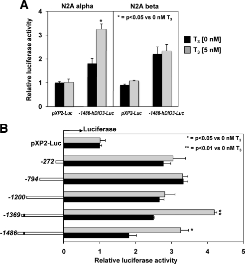
Fig. 4.
T3 response of deletion constructs of the hDIO3 promoter in N2A α and N2A β cells. A, −1486 bp of the hDIO3 promoter-Luc reporter or pXP2-Luc empty vector were transfected into N2A α and N2A β cells and analyzed for luciferase activity after T3 treatment as described in Materials and Methods. Bars represent the mean ± sem of three different experiments, each performed in triplicate. Significant differences between groups are indicated. B, Human DIO3 promoter containing −1486, −1369, −1200, −794, and −272 positions 5′-FR to +72 location at 3′ (just before the native translational start codon) or pXP2-Luc empty vector were transfected into N2A α cells and analyzed for luciferase activity after T3 treatment as described in Materials and Methods. Bars represent the mean ± sem of the three different experiments, each performed in triplicate. Significant differences are indicated. Black bars, T3, 0 nm; shaded bars, T3, 5 nm.

