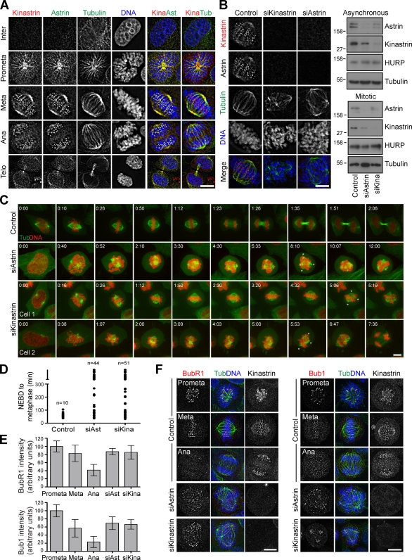
Figure 2.
The astrin–kinastrin complex is required for efficient chromosome alignment and segregation. (A) HeLa S3 cells were stained with antibodies to kinastrin, astrin, and tubulin. DNA was stained with DAPI. (B) HeLa cells were treated with the indicated siRNAs (astrin for 48 h, kinastrin for 72 h) and stained as in A; or cell extracts were prepared from total cells or mitotic shake-offs and Western blotted with the indicated antibodies. Numbers next to gel blots indicate molecular mass in kilodaltons. Bar, 10 µm. (C and D) HeLa cells stably expressing mCherry-histone H2B and GFP–tubulin were treated with control, astrin, or kinastrin siRNA oligos and synchronized with 2.5 mM thymidine 30 h after siRNA addition. After 20 h, the cells were released, incubated for 8 h at 37°C, and then filmed for 12 h. The time required from nuclear envelope breakdown to metaphase alignment was plotted for control, astrin-depleted, and kinastrin-depleted cells (D). (E and F) Control, astrin-depleted, or kinastrin-depleted HeLa cells were fixed and stained with antibodies against kinastrin and BubR1 (left) or Bub1 (right). The staining intensity was quantified and plotted (E). Error bars indicate the standard error of the mean. Bars, 10 µm.

