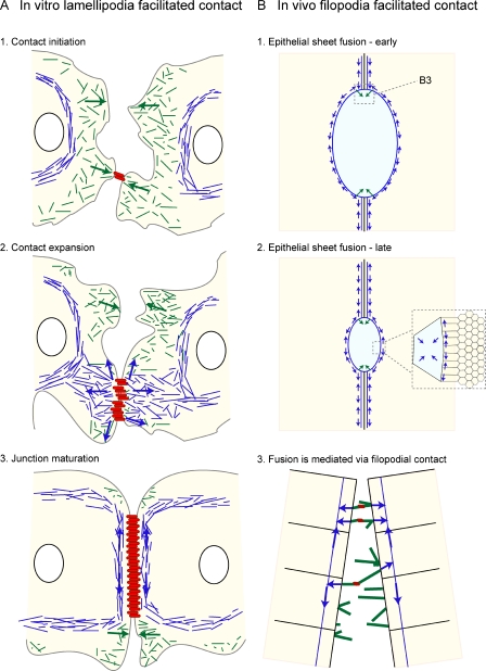
Figure 3.
AJ assembly in vitro and in vivo. (A, 1) Cell contact and E-cadherin engagement in vitro leads to a remodeling of the actin cytoskeleton (green), promoting lamellipodial and filopodial protrusions via Rac, Cdc42, and Arp2/3 activity. (2) These dynamic protrusions promote further E-cadherin interactions and clustering. The nascent AJs are connected to the circumferential actomyosin cable via contractile actin bundles (blue). (3) Myosin-mediated contraction expands intercellular contact and aligns cadherin–catenin complexes (red bars), leading to the maturation of the junction. (B) Fusion between epithelial sheets in vivo again shows cooperation between dynamic protrusions (green arrows) and actomyosin cables (blue arrows). (1 and 2) An actomyosin cable assembles at the edge of each epithelial sheet, forcing the two sheets together. (3) Individual cells on the leading edge of each epithelial sheet form filopodia (green) that engage with one another, forming cadherin–catenin clusters at the points of contact (red), which are required to seal the two sheets together. In the case of the Drosophila embryo during dorsal closure, the ectodermal sheets migrate over a squamous epithelium called the amnioserosa. Here, the apical constriction of amnioserosa cells has been shown to promote dorsal closure (inset). Green arrows represent protrusive activity; blue arrows represent contractile activity.

