Figure 3.
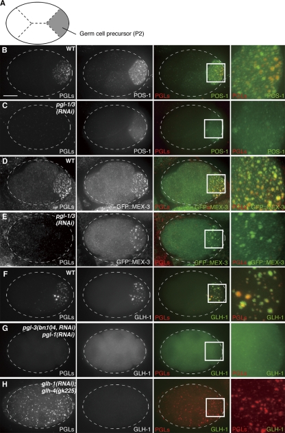
PGL proteins function as the scaffold for P-granule assembly. (A) Schematic representation of a 4-cell stage C. elegans embryo. (B–H) Immunofluorescence images of 4-cell stage C. elegans embryos. Maximum projections of confocal Z-series images that cover whole embryos are shown. In each image, the protein that was detected is indicated. Bar, 10 µm. (B, D, and F) Control wild-type embryos. (C, E, and G) pgl-1(RNAi);pgl-3(RNAi) or pgl-1(RNAi);pgl-3(bn104, RNAi) embryos. (H) glh-1(RNAi);glh-4(gk225) embryo. PGLs: both PGL-1 and PGL-3 detected by the mixtures of monoclonal antibodies KT3 (anti-PGL-3) and KT4 (anti-PGL-1). GFP::PGL-3ΔRGG and GFP::MEX-3 were detected by anti-GFP. POS-1 and GLH-1 were detected by anti-POS-1 and anti-GLH-1, respectively.

