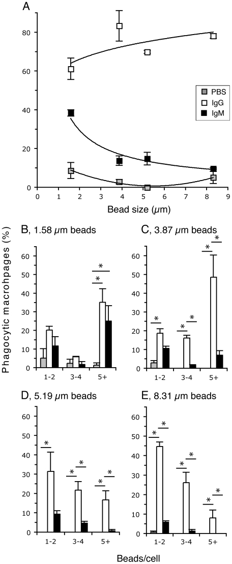
Figure 3. Human IgM preferentially enhances the binding and uptake of smaller-sized particles.
Macrophages were incubated with different size beads that are coated with IgM, IgG or PBS. (A) The percentage of macrophages involved in bead binding and uptake compared to bead size and bead coating condition. Phagocytic macrophages are the cells that contained at least one bead. Non-linear regression analyses between phagocytic microphage (%) and bead diameter: IgG (y = 60.1 x0.1354; r2 = 0.49) and IgM (y = 51.6 x−0.815; r2 = 0.94). These two regression lines are different from zero and to each other (p<0.05). PBS condition does not fit a similar mathematical equation. (B–E) Phagocytic macrophages shown in (A) were examined and counted for the number of beads bound and internalized under varying protein coating conditions and the bead diameter. The percentages of phagocytic macrophages were plotted separately for beads with (B) 1.58 µm, (C) 3.87 µm, (D) 5.19 µm and (E) 8.31 µm in mean diameter. X-axis is the same for charts B–E: 1–2, one to two; 3–4, three to four; 5+, 5 or more beads/cell. Note: Only the phagocytic macrophages are shown in the bar charts; hence, the values for each condition shown in (B–E) will add up to the corresponding values shown in (A); *, p<0.05 (Tukey's test).

