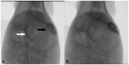
Figure 2. Representative rabbit hepatic angiographic images.
Figure 2A Selective placement of 3F microcatheter (white arrow) and injection of ATONs suspended in lipiodol in the proper left hepatic artery revealed ill-defined hypervascularity tumor staining in the left lobe. Figure 2B Postembolization images show complete stasis of blood flow and demonstrate dense staining of the tumor bed, suggesting successful delivery and excellent distribution of the magnetic nanoparticle fluid mixture in the tumor.

