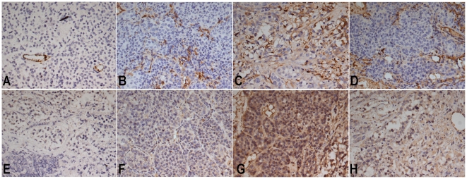
Figure 8. Immunohistochemical staining for CD31 and VEGF.
Representative immunohistochemical staining with CD31 and VEGF monoclonal antibodies in VX2 liver tumors in each group. Microvessels are identified by dark brown (original magnification, 200×). A marked reduction in MVD is observed in the ATON plus hyperthermia group (A) and ATON embolization alone group (B). Abundant microvessels are evident in both the lipiodol TAE group (C) and NS control group (D). Positive VEGFs are recognizable as intensely stained in tumor cell cytoplasm (original magnification, 200×). It is evident that the abundance of VEGF-positive tumor cells is higher in the lipiodol TAE group (G) and NS control group (H) than in the ATON plus hyperthermia group (E) or ATON embolization alone group (F).

