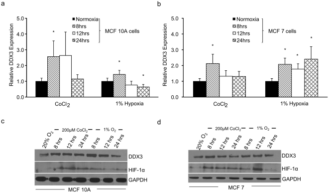
Figure 1. Effect of hypoxic conditions on the expression of the DDX3 gene in breast epithelial cells.
MCF 10A (a & c) and MCF 7 (b & d) cells were cultured under normoxic conditions (20% O2) or subjected to 200 µM CoCl2 or 1% O2 for 8, 12 and 24 h (a & b) qRT-PCR analysis was performed using specific primers for human DDX3 and HPRT as an internal normalization control. The expression level under normoxia was set to 1. DDX3 protein levels (c & d) in cell lysates of MCF 10A and MCF 7 cells were determined by immunoblots using anti-DDX3 antibodies. In these cases, HIF-1α and GAPDH served as controls indicating hypoxic conditions and equal protein loading respectively. Error bars represent ±SD.

