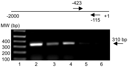
Figure 5. ChIP assay: in vivo binding of HIF-1 to the DDX3 promoter in MCF 10A cells.
At the top of the gel is a schematic representation of the DDX3 promoter. Arrows flank the region (-423 to -115) amplified by PCR with DDX3 promoter specific primers. Gel shows: lane 1- molecular weight (MW) marker, lane 2- total input chromatin, lane 3-acetyl histone H3 precipitation, lane 4-anti-HIF-1α precipitation under hypoxic conditions, lane 5-anti-HIF-1α under normoxic conditions, and lane 6-anti-actin precipitation. Identical volumes from each final precipitation were used for PCR (except for the input chromatin, which was diluted 100x).

