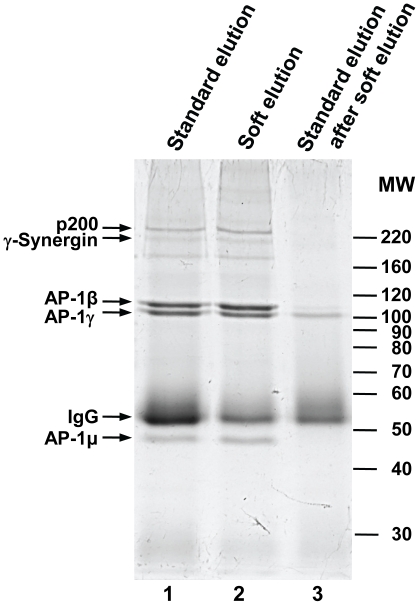
Figure 2. SDS-PAGE analysis of standard and improved elution protocols.
Native immunoprecipitation of the AP-1γ subunit from HeLa cell lysates was performed as described in File S1. Immuno-complexes were recovered by addition of protein A sepharose beads. Prior to elution, beads were split into two equal aliquots. One was subjected to typical high SDS/high heat elution conditions (“standard elution”, lane 1), the other to our “soft” elution protocol (lane 2). Following soft elution, beads were re-eluted using the standard protocol (lane 3), to recover protein still bound to the beads. Thus, lane 3 shows the proportion of immunoglobulin (IgG) that is avoided through soft elution. Proteins in selected bands were identified by mass spectrometry (small arrows). For each band only the top scoring hit is shown. AP-1β, AP-1γ and AP-1µ are components of the AP-1 complex; p200 and γ-Synergin are known AP-1 associated proteins. IgG: rabbit Ig gamma chain C region. Approximate molecular weights are indicated (MW, in kD). Gels were stained with Coomassie G-250.

