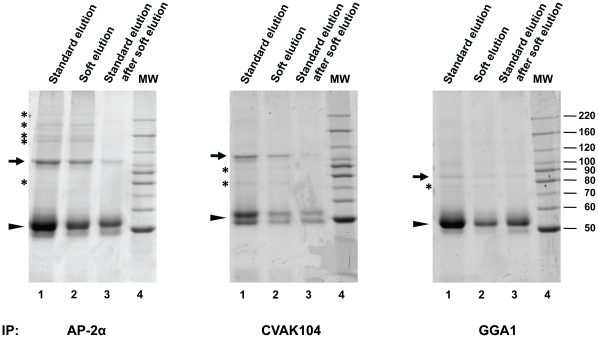
Figure 4. Performance of the improved elution protocol with various rabbit antibodies.
Immunoprecipitations and SDS-PAGE were performed as in Figure 2, with the indicated antibodies (all rabbit polyclonal). Immunoprecipitated proteins were eluted from the protein A sepharose beads using standard conditions (lane 1), or soft-elution (lane 2). Soft-eluted beads were then subjected to standard elution conditions, to recover any remaining material (lane 3). Hence, lane 3 shows the proportion of immunoglobulin (Ig) avoided through soft elution. Lane 4 shows molecular weight markers (MW, in kD). Gels were stained with Coomassie G-250. Small arrows indicate the precipitated primary antigens (identified by molecular weight and through comparison with Western blots). Arrowheads indicate IgG heavy chain bands (identified by molecular weight and abundance). Asterisks indicate examples of proteins that co-precipitate with the primary antigen. The figure shows that soft elution substantially reduces the amount of co-eluting Ig for all tested antibodies, whilst allowing efficient recovery of co-precipitating proteins.

