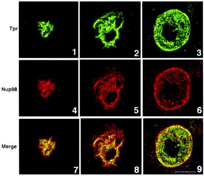
Figure 6.
Colocalization of TPR and Nup98 in an intranuclear network and at the nuclear periphery. HeLa cells were fixed, permeabilized, and incubated with anti-TPR or anti-Nup98 antibodies for double-immunofluorescence analysis by confocal microscopy. Shown are 3 of 32 optical sections across the HeLa cell nucleus in the z axis. Sections 1, 4, and 7 are in a plane above the nucleolus. Sections 2, 5, and 8 are through one pole of the nucleolus; note perinucleolar labeling and spikes that attenuate as they project into the nuclear periphery. Sections 3, 6, and 9 are near equatorial sections through the nucleus; note punctate labeling in the nucleus and the nuclear periphery. (Bar = 10 μm.)

