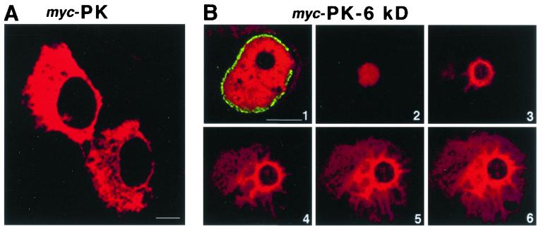
Figure 7.
Immunofluorescence visualization of the TPR network is enhanced by overexpression of a myc-PK-6kDa construct. HeLa cells were transiently transfected with a myc-PK or a myc-PK-6kDa construct. Note that the myc-PK fusion protein is excluded from the nucleus (A), whereas the myc-PK-6kDa fusion protein gains access to the nucleus (B, 1–6). In B, cells were prepared for double-immunofluorescence with anti-Nup358 antibodies and anti-myc antibodies. Six of 64 confocal nuclear sections are shown: an equatorial nuclear section, B1, demonstrates Nup358 staining in green at the cytoplasmic side of the nuclear envelope and the intranuclear staining of myc-PK-6kDa in red. Sections above the nucleolus (B2) and across the nucleolus (B3–6) show the characteristic but much enhanced features of TPR and Nup98 labeling seen in Fig. 6: intense staining of the perinucleolar region from which spikes emanate and attenuate as they project into the nuclear periphery. (Bar = 10 μm.)

