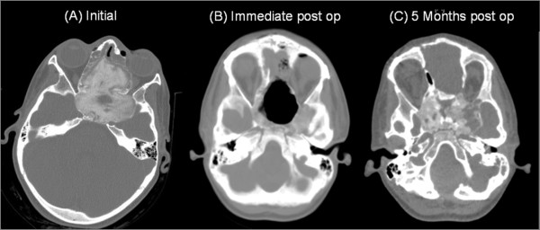
Figure 2.
Series CT scans of the tumor. (A) preoperative CT revealing ground glass appearance is consistent with fibrous dysplasia affecting of the sphenoid bone, ethmoidial bones, LT maxillary sinus, involving the optic foramen and harboring cystic expansible component. (B) Immediate post operative CT scan. (C) 5 months post operative follow up CT scan.

