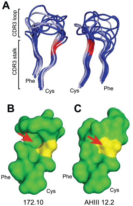
Figure 1. Impact of a G versus S residue at TCRβ position 107.

(A) Three TRBV13-2 (silver) and 7 non-TRBV13-2 (blue) TCRαβ structures (listed in the text) were aligned. Their CDR3β peptidyl backbones are displayed in two orientations, indicating highly conserved stem structures. G107 residues are in pink and S107 in red. The structural impact of a G107 versus S107 is exemplified in the mouse TRBV13-2+ 172.10 TCR (B) and TRBV13-3+ AHIII 12.2 TCR (C). The arrow indicates the location of the S107 side chain in AHIII 12.2 that is absent in the 172.10 TCR.
