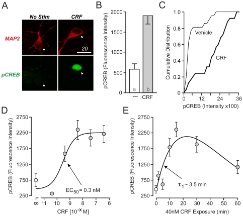
Figure 1. CRF rapidly stimulates CREB phosphorylation.
(A) Immunolabeled confocal images of cultured striatal neurons from 1- to 2-day old rat pups with MAP2 (red) and pCREB (green). Neurons stimulated with CRF (40 nM) for 15 min exhibited increased CREB phosphorylation (Scale Bar = 20 µm). (B) Quantification of immunostaining, revealing CRF-mediated CREB phosphorylation (p<0.0001). (C) CRF induced a rightward shift in the plot of pCREB fluorescence intensity of approximately 80% of striatal neurons (D) CRF increased CREB phosphorylation in a concentration-dependent manner, with EC50 = 0.3 nM. Concentrations ≥ 4 nM induced a signal that differed with statistical significance from vehicle-stimulated (NS) neurons. (E) Time course of 40 nM CRF-induced CREB phosphorylation, with τ = 3.5 min. Statistically different groups are denoted by different alphabetical characters in corresponding bar graphs in this and subsequent figures. P-values <0.05 were considered a priori as significant.

