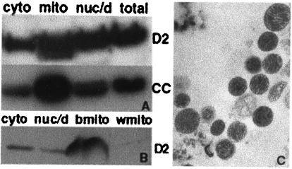
Figure 4.
Distribution of D-AKAP2 in mouse and rat adipose tissue fractions. Mitochondria were isolated from mouse BAT (A) or rat BAT (b) and WAT (w) (B) as described in Experimental Procedures to get the cytosolic supernatant, the nuclear and cell debris pellet and the mitochondria fraction. Then, 50 μg of total protein was loaded and probed with anti-D-AKAP2 (D2) or anti-cytochrome c (CC). Electron microscopy images were collected to indicate the quality of the isolated mitochondria and a representative image of BAT mitochondria is shown in C.

