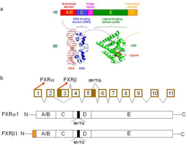
Fig 1. Gene and protein structure of FXR.
(a) General structure of nuclear receptors (NRs). A typical NR contains several domains. The N-terminal region (A/B) contains the ligand-independent AF1 transactivation domain. The C region contains the conserved DNA binding domain. A hinge region (D) connects the DNA- and ligand-binding domain. The E region contains the ligand-binding domain. (b) Schematic diagram of the murine Nr1h4 genomic structure. The mouse Nr1h4 gene is composed of 11 exons and 10 introns. FXRα and FXRβ are transcribed from exon 1 and 3, respectively. A 12 base-pair (bp) insert is located at the 3′ end of exon 5. Alternative splicing from exons 5 and 6 produces two FXR isoforms that either contain the 12-bp insert or not. FXRα and FXRβ have the same amino acid sequence, except for an additional 37 amino acid at the N-terminal of FXRβ. FXRα1 and FXRβ1 have a 4 amino acid insert (MYTG) that is located at the hinge region.

