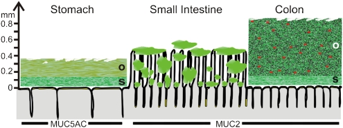
Fig. 1.
Schematic proposal of how mucus is organized in the gut. The thicknesses given are from rat and adapted from the work of Holm and colleagues (13). The red dots in the outer mucus layer of colon illustrate bacteria. The genes encoding the gel-forming mucins (green) expressed by the surface goblet cells in the different parts of the intestine are marked by name. o, outer loose mucus layer; s, inner stratified, firmly attached mucus layer. That the mucus thickness and length of the villi vary along the length of the gut is not illustrated.

